
Scientists Use Neutron Imaging to Reveal Hidden Structures Inside 66-Million-Year-Old T. Rex Bone
 Pecohub
 Scientists have unearthed rare fossilized blood vessels inside the broken rib of Scotty, the largest known Tyrannosaurus rex, using neutron imaging to peek inside the 66-million-year-old bone without damaging it.
The discovery provides a detailed view into how the dinosaur’s body responded to injury before its death and demonstrates how neutron imaging can reveal soft tissues that are rarely preserved in fossils.
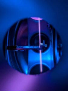
Researchers from the University of Regina in Canada combined neutron imaging with advanced X-ray techniques at the U.S. Department of Energy’s Oak Ridge National Laboratory (ORNL) to create high-resolution 3D images of Scotty’s rib. The procedure allowed them to examine the fossil’s internal structure while leaving the rare preserved tissues intact.
“It’s like winning the lottery,” said Mauricio Barbi, professor of physics at the University of Regina. “Scotty’s rib contains a vast network of mineralized blood vessels that has never before been observed in a fossil.”
Marshy water preserves blood flow
Soft tissues such as blood vessels usually disappear during decay long before fossilization occurs, making Scotty’s rib an extraordinary exception.
According to the researchers, the dinosaur suffered a broken rib during its lifetime. As the injury healed, iron-rich blood flowed into the damaged area and formed new blood vessels to repair the bone.
Before the healing process was complete, however, Scotty died in what scientists believe was a salty marsh. The environment slowed decomposition, allowing the delicate vascular network to survive long enough to become fossilized.
The team said neutron imaging was highly valuable for this study as it is extremely sensitive to light elements, enabling them to spot subtle remnants of preserved soft tissue that are difficult to identify using conventional imaging methods alone.
“Every fossil is a tiny snapshot of the past,” said Jerit Mitchell, a doctoral candidate in physics at the University of Regina who leads the project.
The researchers first detected evidence of fossilized blood vessels in Scotty’s rib in 2020 using micro-computed tomography and synchrotron X-ray imaging at the Canadian Light Source. Those studies confirmed preserved soft tissue and helped scientists examine the healing process on microscopic scales.